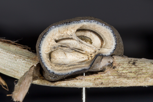
To the extent that they are different from one another, this material may represent either Munkia martyris or Munkia chusqueae (≡Shropshiria chusqueae), both of which are said to be characterized by forming conidia inside of cup-shaped sporodochia, as seen in the macro images. Problematically, the dimensions for both those spp. do not exceed 8mm in any direction, which is significantly smaller than the material seen here. Over the past 130 years, various authors have posited a connection between Munkia and a Mycomalus-like teleomorph, but this has yet to be fully substantiated. Contradictions in the literature regarding conidial morphology and other characters further complicate the study of these often beautiful and widely underappreciated fungi.
Unfortunately, our sections turned up nothing particularly worth photographing, partly on account of the hardness of the tissue (Stevens says it should be “boil[ed] in dilute potash,” which was neither readily on hand in the FLOR herbarium, nor would we have had time to cook the specimen if it were possible to). What’s more, we could locate no conidia; not in any sporodochial cavity we examined, not floating around the medium, not a one. Better luck next time.
We are grateful to MicoLab, particularly Maria Alice Neves and Caue Oliveira, for graciously accommodating our curiosity in these and other Clavicipitaceous specimens.
Substrate: living bamboo culm
Habitat: ?
Ecoregion: Sierra do Mar coastal forests (NT0160)
Collector(s): M.A. Neves
Collection #: MAN 1078
Determination: D. Newman & P. Kaishian


 Danny Newman, some rights reserved (CC BY-NC-SA), uploaded by Danny Newman")