September 20, 2026

Coprinellus Sect. Setulosi Genus Ephemerocybe

Coprinellus Sect. Setulosi Genus Ephemerocybe

What divides up the Coprinellus into the various sections / genus is the veil cells / present: 

Those found in the Core Setulosi clade are mostly smooth, hyaline, thin-walled and scarce, often difficult to notice under the microscope and never visible macroscopically or with
hand lenses,

whereas those in the Micacei/Domestici clades are more rich and macroscopically observable, usually incrusted or have thickened wall.

least reliable character is the occurrence of lageniform cheilocystidia in species with predominantly globose-ellipsoid cystidia.

suggests that spore size and shape can be highly variable in C. congregatus and the C. hiascens group respectively


Phylogeny and species delimitation in the genus Coprinellus with special emphasis on the haired species La´szlo´ G. Nagy Judit Ha´zi Csaba Va´gvo¨ lgyi Tama´s Papp
Mycologia, 104(1), 2012, pp. 254–275. DOI: 10.3852/11-149 

Setulosi clade in the Nagy et al. (2012) phylogeny, is a large group of species with the characteristic setules on the pileus

Section Domestici (Singer) D.J. Schaf. has a veil on the pileus in the form of floccose scales, consisting of chains of fusiform or subglobose cells, often with encrusted walls. In Micacei (Fr.) D.J. Schaf., veil remnants are present in the form of scattered, granulose flocks, often disappearing and consisting of globose cells arising from a matrix of narrow branched hyphae. In Setulosi (J.Lange) D.J. Schaf., the veil may be present or absent, but the pileus and stipe are covered with thin-walled pileocystidia and caulocystidia, respectively

The genus Coprinellus (Basidiomycota; Agaricales) in Pakistan with the description of four new species. MycoKeys 39: 41–61 (2018) doi: 10.3897/mycokeys.39.26743. Shah Hussain1, Muhammad Usman, Najam-ul-Sehar Afshan, Habib Ahmad, Junaid Khan, Abdul Nasir Khalid

Posted on September 20, 2026 10:36 PM by davidwhyte | 0 comments | Leave a comment

September 18, 2026

AI generated polypore key - has not been fact checked.

Google AI generated, take with pinch of salt. 

Macro-Key to Major Genera of Bracket Fungi
1. Fruiting body with a distinct central, eccentric, or lateral stem. → Go to 2
1. Fruiting body without a stem (sessile), attached directly to wood as a shelf or hoof. → Go to 4 [1]

Stem Present
2. Cap surface has a distinct shiny, lacquered, or shellacked appearance.
    2. Cap surface is dull, not lacquered. → Go to 3 [1]
    3. Flesh is pale (white to cream); pore surface does not blacken with age.
    • → Polyporus (e.g., P. squamosus / Cerioporus) [1, 2]
      3. Flesh is brown to dark ochre; cap is velvet-textured and turns black with KOH.

        Stem Absent (Sessile Shelves/Hoofs)
        4. Flesh is soft, fleshy, moist, or watery when young; typically annual. → Go to 5
        4. Flesh is tough, leathery, corky, or rock-hard/woody; annual or perennial. → Go to 7 [1, 2, 3, 4]

        Soft / Fleshy Brackets
        5. Fruiting body grows in large, bright yellow-to-orange overlapping clusters (rosettes).
        • → Laetiporus (Chicken-of-the-Woods) [1, 2]
          5. Fruiting body is single or small overlapping shelves; colors are duller. → Go to 6
          6. Flesh exudes a blood-red juice when squeezed; pore surface darkens when bruised.
          • → Fistulina (Beefsteak Fungus) [1, 2]
            6. Flesh is soft, white, or bruising blue/brown; typically small and growing on conifers.
            • → Postia / Rhodofomes [1, 2]

              Tough, Leathery, or Woody Brackets
              7. Interior flesh is white, cream, or pale pinkish. → Go to 8
              7. Interior flesh is yellow-brown, rusty-brown, or dark brown. → Go to 11 [1, 2]

              Pale-Fleshed, Tough/Woody Brackets
              8. Bracket is thin, leathery, flexible, and heavily zoned with colorful bands on top.
              • → Trametes (e.g., T. versicolor / Turkey Tail) [1, 2]
                8. Bracket is thick, corky, or woody; not flexible. → Go to 9
                9. Pore underside turns dark brown when scratched; cap has a hard, crusty, dull gray/brown shell.
                • → Ganoderma (sessile forms like G. applanatum / Artist's Conk) [1, 2, 3]
                  9. Pore underside does not darken significantly when scratched. → Go to 10
                  10. Perennial hoof-shaped conk; interior flesh is wood-colored with layers of white tubes.
                  • → Fomes (Hoof Fungus) [1, 2, 3]
                    10. Perennial conk with a distinct red-to-orange exterior band along the margin.

                      Brown-Fleshed, Woody Brackets
                      11. Bracket is perennial, extremely hard, and heavily cracked with age; flesh turns black instantly with a drop of KOH.
                        11. Bracket is annual or biannual, softer, or fibrous; often velvety when young. → Go to 12
                        12. Pores are large and distinctly irregular, maze-like, or elongated.
                        • → Gloeophyllum / Daedalea [1, 2]
                          12. Pores are small, circular, and regular; cap surface is hairy or velvety.
                          • Inonotus [1, 2, 3]


                          Genus Growth Form Flesh Color Texture Key Field Feature
                          Ganoderma Large shelf or stalked Brown to Pale Rigid/Woody White pore surface stains brown when scratched
                          Trametes Thin, multi-tiered White Leathery Highly colorful, fuzzy, zoned rings on top
                          Laetiporus Overlapping rosettes White to Yellow Fleshy/Chalky Vibrant sulfur-yellow to orange cap colors
                          Fomes Hoof-shaped Wood-brown Rock-hard Layered, perennial tube structures
                          Phellinus Massive shelf/conk Dark Brown Woody Rimose (deeply cracked) top surface
                          Posted on September 18, 2026 01:50 AM by davidwhyte | 0 comments | Leave a comment

                          September 15, 2026

                          pleurotus / oyster - new species in NZ

                          Copy from paper, showing key differences

                          Multi-gene phylogeny and morphology of Pleurotus in Aotearoa New Zealand reveal a new variety of Pleurotus pulmonarius

                          298 . pulmonarius was originally described from Sweden from fruitbodies associated with
                          399 Birch trees. Hilber (1982) revised the European species of Pleurotus and studied the
                          400 morphology of P. pulmonarius fruitbodies from various host trees. The species does not
                          401 have a modern interpretive epitype. The origin of commercial strains in cultivation in New
                          402 Zealand is mostly unknown but phylogenetic data indicate an Asian origin.
                          403 Collections of P. pulmonarius from indigenous habitats in New Zealand possess some
                          404 consistent morphological differences to fruitbodies derived from imported commercial
                          405 strains, and from Hilber’s concept of the European wild type. In New Zealand fruitbodies
                          406 from the wild, or from ex-wild type strains:

                          the stipe is relatively short
                          the lamellae 407 relatively deep
                          and they run almost the entire stipe length without a significant decrease in depth,
                          and there is no anastomosing between the lamellae at the stipe base.

                          409 Microscopically the

                          spores are relatively large and frequently > 10 μm in length.
                          In some specimens there is abundant germination of spores in situ.

                          Fruitbodies from cultivated
                          411 imported strains and Hilber’s description of European wild-type have relatively long
                          412 stipes, the lamellae terminate well before the stipe base, become shallow, and with clear
                          413 anastomosing between lamellae. Microscopically the spores of P. pulmonarius sens. str.
                          414 are generally shorter and often < 9 μm long. The combined spore data for examined non-
                          415 New Zealand specimens (PDD 124110, 124112 and 124160) are: length 8.7 μm, (σ =
                          416 0.44), width 4.1 μm (σ = 0.23), Q = 2.15 (σ = 0.14), n = 17+20+20.


                          417 In New Zealand P. pulmonarius var. aotearoa may be distinguished from the similarly
                          418 coloured dimitic P. parsonsiae because the latter has a much tougher consistency due to 
                          419 the presence of abundant sclerified hyphae, and the fruitbodiesare often fused at the
                          420 base. P. parsonsiae lamellae are also more narrowly spaced than P. pulmonarius var.
                          421 aotearoa lamellae.


                          422 Although a rare occurrence, P. pulmonarius var. aotearoa has been observed to fruit on
                          423 living trees (iNaturalist #26320495 on Cordyline). This phenomenon is also seen in P.
                          424 australis (PDD 120236, iNaturalist #101389646, iNaturalist #197760600, iNaturalist
                          425 #110440505). Several species within the P. eryngii complex also grow on living plants
                          426 (Jia et al., 2024), but it remains unresolved whether they act as pathogens or grow
                          427 saprotrophically on necrotic plant tissue (Carlavilla & Manjón, 2023). Further research is
                          428 required to determine the lifestyle of P. pulmonarius var. aotearoa and P. australis.

                          Posted on September 15, 2026 01:26 AM by davidwhyte | 0 comments | Leave a comment

                          July 27, 2026

                          toothed fungi in NZ - not spell checked

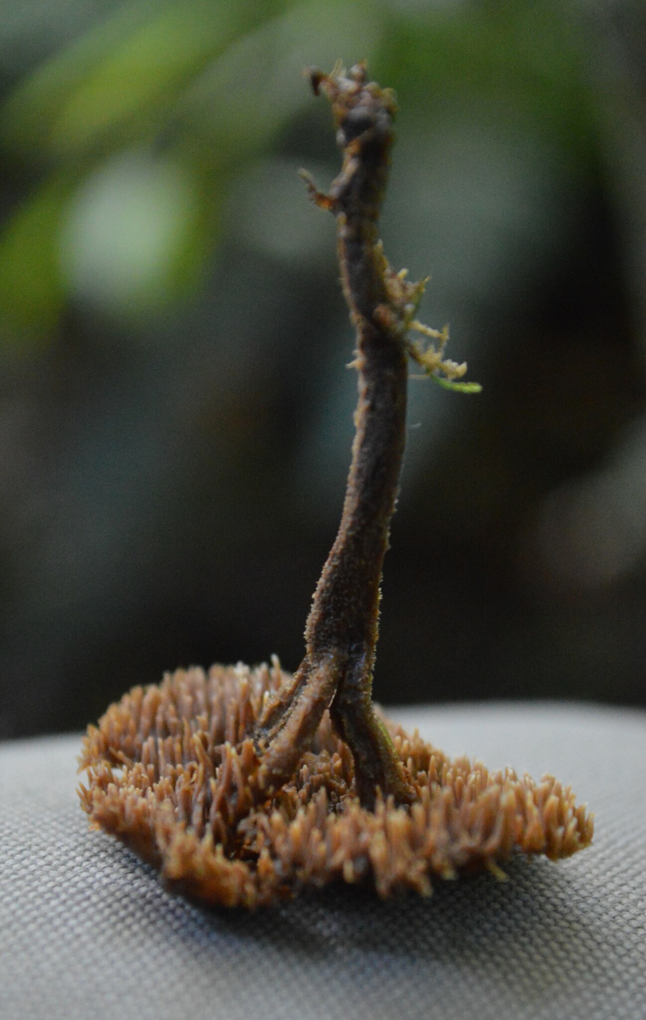
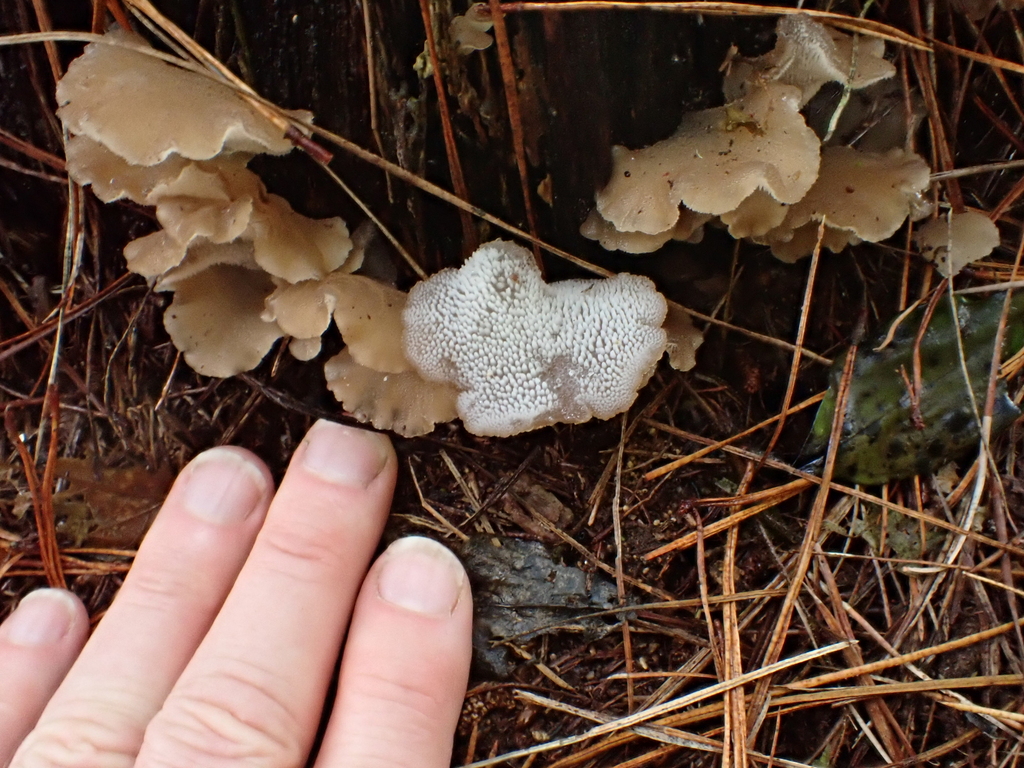
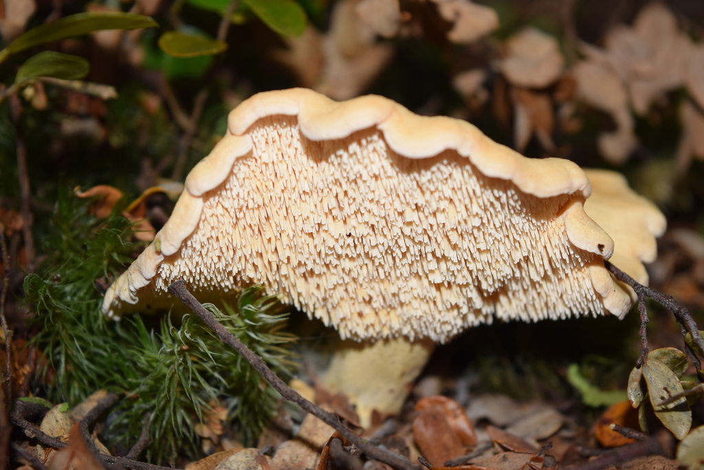
                          Toothed fungi in NZ. There are a number of genus that have toothed fungi. This is a post attempting to give simple defn to help with ID'ing to genus level. 

                           Auriscalpium umbella

                          This is unique in appearing to only have one species in NZ. With beech, not robust. Thin stipe, small cap size, brown tones. 

                          Pseudohydnum

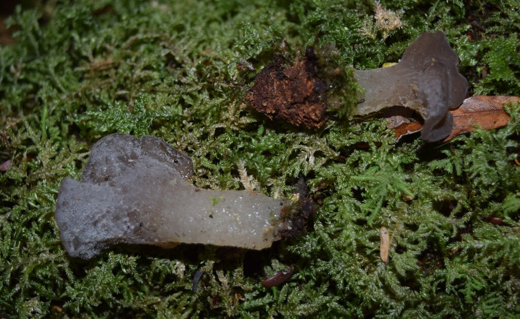
                          Jelly fungi, on wood / decompoers. 

                          Pseudohydnum orbiculare

                          Indigenious and pinus radiata - Clamped, whereas P. viridimontanum is not. Dark tones on cap. 

                          Pseudohydnum totarae

                          Found in broadleaf forest, on wood often well rotted has clear stipe. 

                          Pseudohydnum gelatinosum - NOT recorded in NZ 

                          Pseudohydnum viridimontanum

                          One observation recorded in NZ recorded on tawa. Indicates criptic species overlap with P


                          they key in the describing paper is below. It states that density of spines can tell the two species apart. Not that "shape, colour, spine density to be too variable" to tell species apart. So this cannot be used with 

                          1. Basidiomata stipitate 2

                          1′. Basidiomata sessile or rudimentarily stipitate 3

                          2. Basidiomata confluent; basidiospores 5.5–6.5 × 4.8–5.7 μm P. totarae

                          2′. Basidiomata nonconfluent; basidiospores 6.4–9.7 × 5.4–8.8 μm P. brasiliense

                          3. Pileus surface brown to grayish brown, dark brown, or reddish brown; spines sparse (0.5–1.5 per mm) 4

                          3′. Pileus surface whitish to grayish, grayish yellow, or pale pinkish orange; spines not sparse (≥2 per mm) 5

                          4. Basidiomata sessile; simple hyphidia present P. orbiculare

                          4′. Basidiomata short to rudimentary stipitate; dendrohyphidia present P. brunneovelutinum

                          5. Basidiospores ≥7.2 × 6.0 μm; on Eucalyptus and Nothofagus wood; restricted to Tasmania, Australia P. tasmanicum

                          5′. Basidiospores ≤7.0 × 6.0 μm; on angiosperm wood; Neotropical 6

                          6. Basidiospores subglobose to broadly ellipsoid (Qm = 1.18); inflated segments in spine trama absent; clamp connections present P. cupulisnymphae

                          6′. Basidiospores broadly ellipsoid to ellipsoid (Qm = 1.37); inflated segments cylindrical to fusiform present in spine trama; clamp connections absent P. viridimontanum


                          Genus Hydnum 

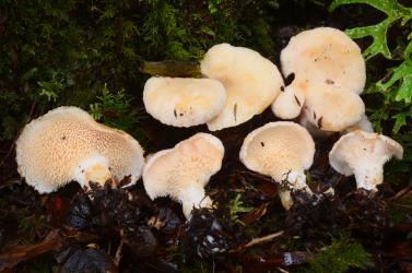
                          Appear bell curve centred July, so fruiting June-Sept which is later than most fungi in NZ. Not jelly fleshed and mycorhizal. Spore illistrations not easy to come by but appear smooth spherical to cylindrical. 

                          Hydnum crocidens
                          Mycohrizal white 

                          Hydnum sp. 'Waima'

                          A cryptic version of H. crocidens, also in Australia

                          Hydnum wellingtonii - This comes from H. crocidens. Var. wellingtonii. Unsure of how to tell apart from H. crocidens 

                          Hydnum sp. 'Nelson Lakes'. - with beech and has dark cap surface

                          Credit: Cooper

                          H.mcnabbianum with Manuka / Kanuka dark cap, also found with beech(?)

                          Credit: Clive Shirley


                          Family Boletopsidaceae has the following genus:

                          See this phyology as things are more complex than the species listed below. Also critical paper is Douch, James K.; Vaughan, Luke J.; Cooper, Jerry A.; Holmes, Gareth D.; Robinson, Richard; Stefani, Franck; Idnurm, Alexander; May, Tom W.: Taxonomic revision of fleshy species of Hydnellum, Neosarcodon , and Sarcodon (Thelephorales) from Australasia . Mycologia: 1-28.

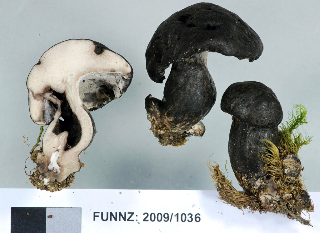
                          The spores of this family are distinct. For example Neosarcoden carbonarius has the following spores

                          Credit : Cooper

                          Genus Boletopsis

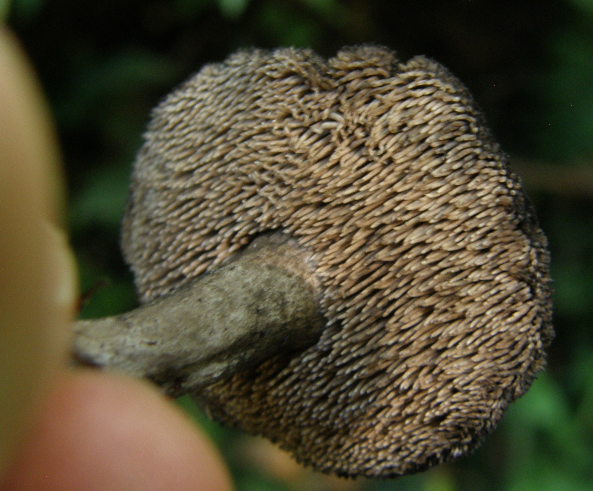
                          Boletopsis nothofagi - this is endagered / only 4 obs, with beech, like a bolete but with tiny spines. At least three species under this name.  
                           
                          Credit: Cooper
                           

                          Genus Hydnellum

                          I have not found this genus (July 26). With beech, bracket like more than mushroom like. However presentation is variable! 
                           
                          Hydnellum pseudoioeides
                           
                           Credit: Cooper 
                           
                          Hydnellum nothofagacearum
                          Named in 2024 
                          Credit: Cooper
                           
                          Hydnellum sp. 'Routeburn
                          Credit: Cooper
                           
                          Credit: Cooper

                          Genus Neosarcodon

                          Neosarcodon carbonarius

                          One would conclude that S carbonarius discussed elsewhere has moved to this name. 'normal' looking mushroom presentation, eg central stipe, domed cap. It is NOT associated with beech but broadleaf / podocarp


                          Genus Sarcodon

                          I have not found any (July 26), no species ID'ed on inat. Biotanz indicates there is one known species in NZ, called Sarcodon sp1. With beech

                          Sarcodon carbonarius'

                          This is in BiotaNZ but notes indicated that the species collected in NZ may not be S. carbonarius and/or S. carbonarius as a species defintion has issues. I would conclude it has moved to Neosarcodon. 


                          In the Northern Hemisphere (or elsewhere, just not recorded in NZ) there is the Bankera genus eg Bankera fuligineoalba. This may be found with exotic trees if present in NZ.  

                          Posted on July 27, 2026 09:23 AM by davidwhyte | 0 comments | Leave a comment

                          June 27, 2026

                          Notes on Waxcaps and Allies Family Hygrophoraceae

                          Copied from various places, attempting to put into one place for ease of access. Not a key, nor definitive, a place to start / things to check.

                          Gliophorus lilacinoides - multicolored and includes lilac
                          a member of Waxcaps and Allies Family Hygrophoraceae
                          https://www.inaturalist.nz/observations/286218398

                          Comments by Jerry post DNA testing:

                          With these Gliophorus you need to get a photo of the gill attachment and gill edge if you can. In this case, from the dried specimen, they are narrowly attached and there is no sterile thread on the gill edge. So you are in the viridis/lilacinoides/ostrinus group. Separating viridis is easy but ostrinus/lilacinoides not so much. The colours (reddish pink versus purple), and changes in colour with age are variable (the sequenced specimens of the same thing tell you that), so colours are not reliable. It comes down to Horak's stated difference in spore size, and as often the case with Horak's species you find G. ostrinus was based on a single collection, so it may not be typical, in either colour or spore size. If you look at the tree ...
                          https://www.funnz.org.nz/sites/default/files/2024JulMultiHygrophoraceae_1.pdf
                          The lilacinoides PDD specimens are Clive's and he didn't provide spore measurements - and I haven't looked. All collections I've measured have larger spores (for this specimen they are 8.5 x 4.2.) and suggest lilacinoides. I think the name G. ostrinus should be ignored and we have 4 species that fit Horak's G. lilacinoides. Maybe there are obvious differences, but I don't see them. This specimen is the same as jac16848 and your https://inaturalist.nz/observations/285558926

                          Dark brown cap and white gills - check Cuphophyllus griseorufescens
                          Dark cap and purple underside check Hygrocybe keithgeorgei
                          Yellow stipe, purple underside and brown-yellow ish cap - Hygrocybe lilaceolamellata
                          Brown-red -purple cap and viscid - Humidicutis multicolor
                          Yellow-white decurrent gills - Cuphophyllus salmonipes

                          Posted on June 27, 2026 06:36 AM by davidwhyte | 6 comments | Leave a comment

                          June 25, 2026

                          Boletes with exciting colors found with manuka / kanuka

                          Notes, still not fully digested. Bolded for clear differences

                          Yellow spored with red tones boletes with manuka or kanuka


                          Boletus novae-zelandiae

                          SPORES: spore print not obtained; spores melleous, elliptic-subfusiform, 14.2-18.2 X 4.5-5.5 µm, smooth.
                          HYMENIUM: basidia hyaline, clavate, 26-36 X 8.5-10.5 µm, 4-spored;
                          cystidia scattered, hyaline, thin-walled, subcylindrical to cylindrical, 36-55 X 4.5-6.5 µm..
                          HYMENOPHORAL TRAMA: bilateral of the Boletus subtype;
                          clamp connections absent.
                          ONTEXT OF PILEUS: yellow, unchanging or becoming faintly red on exposure to air.
                          SMELL: not distinctive.

                          Boletus rawlingsii

                          SPORES: spore print insufficient for accurate determination, but near olive brown; spores melleous, broadly elliptic-subfusiform, occasionally obovate, ll-13.8-(14.9) X 4.5-5.9-(6.9) µm, smooth.
                          HYMENIUM: basidia hyaline, clavate, 35-49 X 9.5-11.5 µm, 4-spored;
                          cystidia numerous, scattered, hyaline, thin-walled, ventricose-rostrate, 42-72 X 8.4-11.5 µm.
                          HYMENOPHORAL TRAMA: bilateral of the Boletus subtype; clamp connections absent.
                          CONTEXT OF PILEUS: pallid yellow to yellow, unchanging on exposure to air.
                          TASTE: unpleasant, slightly acrid.
                          SMELL: not distinctive.
                          CHEMICAL REACTIONS: KOH on pileus—darkening; on context of pileus—reddish; NH4OH on pileus and context—no reaction.

                          Boletus leptospermi

                          SPORES: spore print khaki (Tawny-Olive); spores melleous, broadly elliptic-subfusiform, 8.4-10.8-01.5) X 4-5 µm, smooth.
                          HYMENIUM: basidia hyaline, clavate, 27-42 X 8-10.8 µm, 4-spored;
                          cystidia numerous, scattered, hyaline or brownish, obclavate to narrowly ventricose-rostrate, 39-60 X 6-11.5 µm.
                          HYMENOPHORAL TRAMA: bilateral, of a modified Boletus subtype, mediostratum strongly pigmented; clamp connections absent.
                          CONTEXT OF PILEUS: pallid lemon yellow, rapidly bluing on exposure to air, occasionally with red tints around larvae tunnels.
                          TASTE AND SMELL: not distinctive.
                          CHEMICAL REACTIONS: KOH on pileus and context of pileus—no reaction; NH4OH on pileus—no reaction; on context—blue.


                          Xerocomus rufostipitatus

                          SPORES: spore print not obtained; spores pallid melleous, elliptic-subfusiform, 9.8-12.6-(13.6) X 3.5-4.9 µm, smooth.
                          HYMENIUM: basidia hyaline, clavate, 28-39 x 7.5-11.5 µm, 4-spored;
                          cystidia scattered, hyaline thin-walled, subcylindrical to narrowly ventricose-rostrate, occasionally 1-septate, 38-58 X 7-9 µm.
                          HYMENOPHORAL TRAMA: bilateral, of the Phylloporus subtype;
                          clamp connections absent.
                          CONTEXT OF PILEUS: cream, unchanging on exposure to air.

                          Xerocomus griseoolivaceus

                          SPORES: spore print olive brown (Isabella Color); spores pallid melleous, elliptic-subfusiform, 8.4-10.4-(11) x 3.9-4.5 µm., smooth.
                          HYMENIUM: basidia hyaline, clavate, 25-33.5 X 7-9-8 µm, 4-spored;
                          cystidia sparse, scattered, hyaline, thin-walled, ventricose-rostrate, 42-60 x 8-11.5 µm.
                          HYMENOPHORAL TRAMA: bilateral, of the Phylloporus subtype; clamp connections absent.
                          CONTEXT OF PILEUS: sordid white, unchanging on exposure to air.
                          TASTE AND SMELL: not distinctive.
                          CHEMICAL REACTIONS: KOH and NH4OH on pileus and context of pileus—no definite reaction.


                          Posted on June 25, 2026 07:48 AM by davidwhyte | 0 comments | Leave a comment

                          Stephanospora

                          Stephanospora - have four known species in NZ. These consentrate in the North Island so likely cryptic species present. 

                          Stephanospora aorangi. Habit and Habitat: solitary or in small clusters. In New Zealand, epigeous under loose leaf litter in podocarp-broadleaf
                          forest or mature tea-tree scrub. Remarks: This species co-occurs, in both New Zealand and Australia, in the same habitats as several other new species, but is readily distinguished by its larger, broadly ellipsoid spores with much larger spines and corona.

                          Stephanospora kanuka. Stephanospora kanuka includes collections with predominantly either 2- or 4-spored basidia within a sporocarp, and is distributed in the northern central and west coast of the North Island of New Zealand, where it can occur in some of the same habitats and sites as S. poropingao. Stephanospora kanuka differs in the presence of a faint sweet odour,
                          spores that are more subglobose to ellipsoid, with ornamentation tending to be more spiny and irregular in height, thus ‘messier’ in appearance. This species is predominantly found in association with kanuka, whereas S. poropingao is predominantly found in podocarp-broadleaf forest. However, kanuka can occur in gaps within podocarp-broadleaf forest so examination of spores is required to confirm the identification of these two taxa. The number of spores affects spore size, in that bispored basidia tend to have slightly larger spores (within 0.5 mm). Stephanospora kanuka belongs to subclade A(ii), and is sister to S. pounamu from the South Island, which has smaller spores with lower ornamentation, and also an association with mixed broadleaf-podocarp forest with scattered kanuka.

                          Stephanospora poropingao. Remarks: Stephanospora poropingao may be distinguished from the co-distributed S. kanuka, in the more globose spores, with more robust spore ornamentation with more wedges and short ridges, and association with podocarp-broadleaf vegetation. The majority of collections of Stephanospora poropingao are from the north-west coast of the North Island of New Zealand. 

                          Stephanospora pounamu. Stephanospora pounamu is endemic to one small region of the northern tip of the South Island, in the Nelson area. The bright colour, tendency to fruit at the soil surface, and extensive collecting of fungi throughout the South Island by numerous mycologists, means this taxon should have been collected if it were present elsewhere. Stephanospora pounamu may be differentiated by the subcutis of large inflated cells to 35e60 mm broad; globose to subglobose spores and low ornamentation. 


                          Frustratingly the PDF doesn't seem able to copy out the key text. So see paper for fuller key

                          Large prominent corona (large pyramidal spines with a prominent, circular, basal crown or corona) , spore orgamentation 2 - 6 microns - S. aorangi Spores below

                          Spores with small less visable corone and spore ornamentation 0.1-3 microns - 2


                          Spores globose to subglobose - 3 

                          Spores subglbose to ellipsoid - associated with Kamuka / Manuka North Island S. kanuka  - spores below

                          Definitions Q  
                          Globose 1-1.05
                          Subglobose 1.05 – 1.15
                          Broadly ellipsoid 1.15 – 1.3
                          Ellipsoid 1.3 – 1.6


                          3 Spores 10-14 x 10-13 - inside pale yellow ith olive tones, ornamentation of robust wedge spines or warts in short ridges 1 -2 microns tall, corona 5-7 x 1-3 microns broadleaf podocarp North Island S. poropingao  - spores below 


                          3' Spores 8-11 x 8-11 - odour none, inside pale orange-brown broadleaf podcarp - South Island S. pounamu - spores below


                          Cryptic diversity in the sequestrate genus Stephanospora (Stephanosporaceae: Agaricales) in Australasia link

                          http://dx.doi.org/10.1016/j.funbio.2014.12.007

                          Posted on June 25, 2026 05:06 AM by davidwhyte | 1 observation | 0 comments | Leave a comment

                          June 24, 2026

                          Dark capped mycena

                          Notes produced from reading cooper's preliminary key on mycena Mycena sp. 'Ahuriri Reserve (PDD 80918)' has a note to also compared with M. leptocephala , atroavellanea cf, and M. morris-jonesii since these might visually be similar at a macro level. Below are my notes on how to tell apart:


                          Mycena sp. 'Ahuriri Reserve (PDD 80918)'. CC's distinct. 

                          Mycena leptocephala -  nitrous bonnet. Thicker CC's more fat fingers than long thin neck 


                          Mycena morrisjonesii - cap elements not dextroid (or semi-dextroid) CC's filled with a reddish cell sap (can't see in photos)

                          AI - "A key identifying feature is that they are filled with a distinct reddish plasmatic pigment and measure about 50-70 μm × 10-15 μm:"


                          Mycena atroavellanea - more common and has dextroid cap cells 

                          AI - CC's "25 µm × 8 µm to 40 µm × 13 µm in size"

                          ---

                          So measuring CC size could given an indication of > 40 μm size is found. However only small CC doesn't not rule out M. morrisjonesii. 

                          Posted on June 24, 2026 09:37 AM by davidwhyte | 1 observation | 0 comments | Leave a comment

                          June 23, 2026

                          Hypholoma notes

                          There are three hypholoma that I struggle to tell apart. Attempted to find chelio and pesudo chelio (can't remember word) which are nipple like cells didn't appear to be that different.

                          Hypholoma sp. 'Totaranui - doesn't have the micro work done, although it is likely it is very similar to previously as H. subviride cf.

                          Nipples are only present on Hypholoma acutum, they are distinct and very pointy. Does not mean that they all have these nipples. So some have no nipples

                          Posted on June 23, 2026 12:50 AM by davidwhyte | 1 observation | 0 comments | Leave a comment

                          June 21, 2026

                          Pseudohydnum in NZ descriptions / key etc


                          Pseudohydnum-orbiculare used to be ID'abel compared to the pseudohydnum-totarae. I'm unsure of how, but suspect enviroment / substrate / size / macro structure would be used. However since Pseudohydnum viridimontanum has been found in NZ in 2025, this indicates that micro work is required. 


                          Pseudohydnum orbiculare J.A. Cooper, Type—New Zealand. South Island, West Coast Region, Ship Creek, Haast, on an unidentified rotten branch,

                          Etymology—Orbiculare (Lat.): refers to the species having orbicular basidiomata.

                          Diagnosis—Differs from other Pseudohydnum species by pileate basidiomata without a well-formed pseudostipe, orbicular pilei, dark-gray to black pileal surface when fresh, hymenophore with sparse spines (0.5–1 per mm at base), growth mostly on angiosperm, and distribution so far restricted to New Zealand. 

                          Basidiomata—Annual, gelatinous when fresh, pileate, not confluent, in grouped tiers. Pilei laterally attached, with a narrow point of attachment, orbicular to dimidiate, up to 50 mm diam., pileal surface white to grayish brown (7E3–9E3) to reddish brown (11E5), smooth to minutely velutinate, with scattered, bluntly conical spines up to 0.2 mm long.  Hymenophore white to pale-brown (8B2). Hymenophore spines white, conical, gelatinous, 2–4 mm long, and 1–2 mm wide at the base, 0.5–1 per mm at base. Context 0.5–1.5 mm thick, concolorous with pileal surface. 


                          Hyphal structure—Monomitic; generative hyphae with clamp connections. Contex-tual and spine tramal hyphae hyaline, thin- to slightly thick-walled, frequently branched, agglutinate,1.5–9 µm in diam. Basidia longitudinally cruciate-septate, 10–14 × 10 µm, with four sterigmata up to 10 × 2 µm. Probasidia lageniform to clavate with stalks 1.5–2 µm in diam. Hyphidia irregular, mostly unbranched, slightly swollen towards apex, clamped at the base. Basidiospores (excluding apiculus) broadly ellipsoid to subglobose, hyaline, thin- walled, occasionally with a guttule, germinating by repetition,  6.5–7.9 × 5.6–6.8 µm,

                          Distribution—Distributed throughout New Zealand but perhaps more common in the south. 

                          Habitat—The species is commonly associated with angiosperm hosts, including southern beech (Nothofagaceae), but several collections were obtained from rotten trunks of Pinus Radiata

                          Figure 3. Basidiomata and microscopic structures of Pseudohydnum orbiculare. (A) Basidiomata (holo- otype PDD 112653). (B) Basidiomata (PDD 112654). (C) A hyphidium. (D) Basidia. (E) Basidiospores. type PDD 112653). (B) Basidiomata (PDD 112654). (C) A hyphidium. (D) Basidia. (E) Basidiospores.


                          Pseudohydnum viridimontanum 

                          Diagnosis: Pseudohydnum viridimontanum is similar to P. cupulisnymphae but can be distinguished by its smaller and mostly short-stipitate basidiomata, smaller basidiospores (5.8–6.9 × 4.4–5.0 μm), presence of cylindrical to fusiform inflated elements in the spine trama, and absence of clamp connections. In addition, it was only found on Vernonanthura discolor wood.

                          Etymology: viridis (Latin) = green, montanum (Latin) = hill, mount, mountain; referring to the type locality, i.e., “Monte Verde” in Portuguese, which means Green Hill.

                          Morphology: Basidiomata small-sized, gelatinous when fresh, pileate, laterally stipitate or pseudostipitate, solitary or in imbricate clusters, at first as a globose to bell-shaped primordia, then expanding to orbicular, flabelliform or reniform. Pileus up to 35 mm diam, surface smooth to minutely rough, white (1A1) at first, then chalk white to grayish white (1B1 to somewhat paler) or grayish yellow (1B3, 3B2); margin sharp, even to crenate, sometimes undulating. Hymenophore white (1A1) to whitish, hydnoid. Hymenophore spines sharp-tipped, up to 0.8 mm long, 2.5–4 per mm, white (1A1) to translucent white. Stipe short to rudimentary. Context concolorous with the pileus, translucent appearance when wet, thick centrally and gradually thinner toward the margin, unchanging. Odor mild. Taste mild or pleasant.

                          Hyphal system monomitic, generative hyphae clampless, hyphae unchanged in KOH. Pileal surface subtrichodermial to trichodermial, composed of scattered fascicles of cylindrical hyphae (2.5–5.2 µm diam) that are colorless in mass and frequently septate, thin- to slightly thick-walled; terminal elements poorly differentiated.
                          Contextual hyphae thin- to slightly thick-walled, hyaline, interwoven, frequently branched, commonly anastomosing, 1.5–5.0 diam. Spine tramal hyphae thin-walled toward the tips of the spines, slightly thick-walled toward the core, smooth, hyaline, branching, interwoven, 1.0–3.0 µm diam, sometimes forming intercalary and terminal inflated segments; intercalary inflated segments cylindrical to fusiform, 68–80 × 6.9–13.0 µm; terminal inflated segments long clavate, 40–85 × 9.0–14.0 µm. Hyphidia simple, thin-walled, hyaline, 1.0–2.7 µm diam at the apex, covering basidial cells.
                          Basidia 4-celled, longitudinally septate, multigutullate, barrel-shaped to subglobose, 9.2–12.0 × 8.1–10.6 µm; sterigmata up to 22 × 1.4–2.3 µm, mostly tubiform, usually slightly flaring at apex. Probasidia subglobose to short clavate, 8.8–9.0 × 6.5–7.5 µm, mostly multigutullate.
                          Basidiospores [90/3/2] (5.0–)5.6–6.9(–7.3) × (3.9–)4.3–5.0(–5.5) μm [Lm = 6,2–6.5 μm; Wm = 4.6–4.7 μm; Q = (1.16–)1.23–1.53(–1.66); Qm = 1.32–1.37], broadly ellipsoid to ellipsoid, rarely elongate, nonamyloid, nondextrinoid, acyanophilous, hyaline, thin-walled, mostly germinating by repetition, sometimes by a germ tube conspicuously long, up to 28 μm long.

                          Habitat and distribution: Growing on decayed wood of Vernonanthura discolor in upper montane mixed ombrophilous forest of Southeastern Brazil, at 1600 m a.s.l. Only known for the Brazilian state of Minas Gerais.

                          Notes: In the field, P. viridimontanum immediately evokes P. cupulisnymphae because of the white to grayish yellow pileus and occasional basidiomata with a rudimentary stipe. Both species also display similar hymenial to subhymenial anatomy. However, P. cupulisnymphae differs by its stouter and mostly sessile basidiomata, larger basidiospores (5.8–8 × 5.1–7 μm) with a lower Qm value (Qm = 1.10–1.18), clamped generative hyphae, and the absence of cylindrical to fusiform inflated segments in spine trama. Furthermore, P. viridimontanum is also different by the ontogenetic configuration in young basidiomata because at first it forms a globose to bell-shaped basidiome that rapidly expands and assumes a mature flabelliform shape. In contrast, all young stages observed throughout P. cupulisnymphae collections involve a distinctly funnel-shaped or cupulate configuration.

                          Figure 12. Pseudohydnum viridimontanum (holotype; NCC266, FIFUNGI292). A. Primordia and young basidiomata. B. Mature basidiomata, top view. C. Mature basidiomata, underside view. D. Hymenophoral spines detail. E. Basidia. F. Basidiospores. G. Terminal inflated elements from spine trama. H. Hyphidia. Bars: A–C = 10 mm; D = 5 μm; E–G = 10 μm.

                          Pseudohydnum brunneovelutinum (holotype; AGS118/2022, FIFUNGI283). A–B. Basidiomata. C–D. Detail of the hymenophoral spines. Bars: 10 mm. Photographs: Alexandre G. dos Santos e Silva-Filho.

                          ...Remarkably, accurate analyses of all P. viridimontanum basidiomata failed to spot any clamp connections, 

                          DICHOTOMOUS KEY FOR THE SEVEN CURRENTLY KNOWN TAXA OF PSEUDOHYDNUM FROM THE SOUTHERN HEMISPHERE

                          Confirmed NZ species highlighted in bold. It is likely we have other species in NZ.  

                          • 1. Basidiomata stipitate 2

                          • 1′. Basidiomata sessile or rudimentarily stipitate 3

                          • 2. Basidiomata confluent; basidiospores 5.5–6.5 × 4.8–5.7 μm P. totarae

                          • 2′. Basidiomata nonconfluent; basidiospores 6.4–9.7 × 5.4–8.8 μm P. brasiliense

                          • 3. Pileus surface brown to grayish brown, dark brown, or reddish brown; spines sparse (0.5–1.5 per mm) 4

                          • 3′. Pileus surface whitish to grayish, grayish yellow, or pale pinkish orange; spines not sparse (≥2 per mm) 5

                          • 4. Basidiomata sessile; simple hyphidia present P. orbiculare

                          • 4′. Basidiomata short to rudimentary stipitate; dendrohyphidia present P. brunneovelutinum

                          • 5. Basidiospores ≥7.2 × 6.0 μm; on Eucalyptus and Nothofagus wood; restricted to Tasmania, Australia P. tasmanicum

                          • 5′. Basidiospores ≤7.0 × 6.0 μm; on angiosperm wood; Neotropical 6

                          • 6. Basidiospores subglobose to broadly ellipsoid (Qm = 1.18); inflated segments in spine trama absent; clamp connections present P. cupulisnymphae

                          • 6′. Basidiospores broadly ellipsoid to ellipsoid (Qm = 1.37); inflated segments cylindrical to fusiform present in spine trama; clamp connections absent P. viridimontanum


                          So P. totarae has basidiomata stipiate (which means??) vs sessile or reimentarily stipate 

                          Spines spares and 0.5 - 1.5 per mm is P. orbiculare whereas spines > 2 per mm is P. viridimontanum


                          Other differences:

                          Clamped (P. orbiculare) vs not clamped (P. viridimontanum)

                           6.5–7.9 × 5.6–6.8 q= 1.17 (P. orbiculare) vs 5.6–6.9 × 4.3–5.0 Q = 1.37 (P. viridimontanum)


                          Pseudohydnum gelatinosum - is not found in NZ even though it is on inat. 



                          Posted on June 21, 2026 10:13 PM by davidwhyte | 1 observation | 0 comments | Leave a comment