 Annotations
Jan Jurníček
Joined: Sep 29, 2024 Last Active: Sep 16, 2026 iNaturalist
Now retired, I work as an independent researcher dedicated to the study and microphotography of Rotifera and Cladocera across various aquatic habitats in the Czech Republic. As my focus is primarily on microphotography rather than expert taxonomy, I highly welcome and appreciate any help from the community with precise species identification.
All observations and photographs uploaded here are my own original work. I personally collect all samples, perform the microscopic analysis, and document exact dates and geographic locations. I do not use images from the Rotifer World Catalog or any other third-party sources; I am the sole author and copyright holder of all media shared here.
My Microscopy Setup & Workflow
Hardware:
- Microscope: Euromex BS.1153-PLi trinocular (Brightfield)
-
Objectives:
- Euromex Plan PLi 4x/0.10 ∞/-
- Euromex Plan PLi 10x/0.25 ∞/-
- Euromex Plan PLi 20x/0.40 ∞/0.17
- Euromex Plan PLi 40x/0.65 ∞/0.17
- PLAN 100x/1.25 Oil immersion ∞/0.17
- Camera: ZZCAT SCMOS 05100KPB 5MP USB eyepiece camera mounted via a 0.5x reduction lens.
Software:
ImageView:
The camera's firmware and the bundled ImageView software fully support Flat Field Correction (FFC). To achieve a uniform background, my routine workflow involves using the mechanical stage to move the glass slide away from myself along the Y-axis until the slide is completely outside the objective's field of view. When not using an immersion objective and unless the background is heavily stained, this method allows me to capture and export a pristine reference image of the clear illumination path without altering the vertical focus (Z-axis). This corrected profile accounts for uneven sensor sensitivity and illumination shading (vignetting), and the exported reference can be reused for subsequent sessions as long as the microscope head alignment remains unchanged.Custom Image Processing Software: My own custom-coded application designed specifically for manual focus stacking, sequence manipulation, and background plate composition.
Image Processing & Focus Stacking
Illumination & Aperture Calibration Protocol:
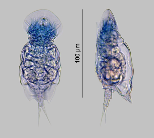
To optimize resolution and maximize the signal-to-noise ratio (SNR) for 20x and 40x objectives, I minimize the microscope illumination, enable "Auto Exposure," and adjust the condenser aperture diaphragm to achieve an exposure rate of 100 fps with an empirically determined sensor gain between 1500 and 1600 (this range is subject to future optimization based on ongoing testing). Next, I increase the illumination intensity until the Auto Exposure forces the camera gain down to its native minimum value of 100. Once the specimen is correctly positioned, I execute Flat Field Correction (FFC) and White Balance using a Region of Interest (ROI) before capturing the focal series by gradually moving the objective away from the specimen.Image Acquisition & Stacking: Due to the extremely shallow depth of field at high magnifications, I photograph the organism (e.g., a rotifer immobilized in glycerin) at multiple focal planes. To process these, I perform manual focus stacking using a custom graphic software that I coded myself to achieve the cleanest and most accurate composite images of key morphological features.
Additionally, my custom software allows me to seamlessly combine focus stacking with multi-shot panoramic stitching. By moving the specimen along the X and Y axes using the microscope’s mechanical stage—while maintaining a perfectly fixed rotation—I can stitch multiple overlapping fields of view together. This advanced capability enables me to generate high-resolution composite images of specimens that are significantly larger than the camera’s native field of view.
Depending on the specimen's position, I use manual focus stacking from bottom-to-top to capture the top view. If needed and possible, stacking from top-to-bottom and mirror-reversing the sequence provides the bottom view.Background Cleaning & Optimization: The software matches the background of all captured views to a tone customizable tone of my choice (such as LightGray). Any background areas that carry no diagnostic information for species identification are replaced with this solid color. This significantly optimizes the JPEG compression and reduces file size without losing essential data. Please note that while the background is digitally optimized for clarity and compression, the specimen itself remains strictly unaltered, preserving all authentic diagnostic morphological features for valid species identification.
Composite & Final Editing: The selected views are arranged into a single composite image (plate) against a solid background. Finally, I expand the histogram and fine-tune the contrast, saturation, and color tone to make the diagnostic features stand out.
External Gallery
My BioLib.cz gallery: biolib.cz/galleryuser/?uid=3747
Following 0 People
jjurnicek is not following anyone.