Species Identified:
Odontella aurita (Lyngbye) C.Agardh, 1832
Genus: Odontella (Lyngbye) C.Agardh
Taxonomic notes on the genera: Odontella
Phyllum: Bacillariophyta
Subphylum: Bacillariophytina
Class: Mediophyceae
Subclass: Thalassiosirophycidae
Order: Eupodiscales
Family: Odontellaceae
Genus: Odontella (Guiry and Guiry 2024)
Type species, synonym(s), etc.: Holotype species: Odontella aurita (Lyngbye) C.Agardh (homotypic synonym: Biddulphia aurita (Lyngbye) Brebisson 1838)
Genus summary:
Similar-looking diatoms of the Eupodiscaceae (e.g. Hobanellia, Odontella, Trieres, Zygoceros) and Biddulphiaceae (e.g. Biddulphia) can be approximately divided, morphologically and genetically, into two broad classifications: 1) ocellate and pseudocellate diatoms, respectively. The ocellus-bearing taxa (Eupodiscaceae) are monophyletic, and thus the ocellus is a useful morphological character in establishing the order Eupodiscaceae. However, the Biddulphiaceae are polyphyletic and dispersed across various lineages of multipolar non-pennate diatoms with a taxonomically confused history (Hoban 1983, Round et al. 1990, Ashworth et al. 2013). Previously, Biddulphioid diatoms included most of the Odontella species (Cupp 1943, Shim 1976, Tynni, R. 1986, Waters et al. 1992), including the taxa found in the Salish Sea; there still exists confusion regarding the naming of these two genera, partly because of the morphological variability of species such as O. aurita (Cupp 1943: 616-163). However, in the last twenty years, with molecular and SEM investigations, the taxonomy of ocelli- and pseudocelli-bearing diatoms and the taxonomy of Odontella have become less ambiguous (Lavigne et al. 2015, Sims et al. 2018, Ashworth et al. 2013). See a detailed genus description in Guiry and Guiry (2024).
Odontella is cosmopolitan in the marine littoral, planktonic, epiphytic and benthic habitats. Both O. aurita (McIntire and Overton 1971, Tynni, R. 1986, Sancetta and Calvert 1988: Odontella longicruris, Waters et al. 1992, Pienitz et al. 2003 and O. obtusa (Rao and Levin 1976, Shim 1976) appear to be common diatoms within the Salish Sea and along the west coast of North America (Cupp 1943). Hobaniella longicruris (Greville) P.A.Sims & D.M.Williams 2018 (formerly Odontella longicruris), with O. aurita and O. obtusa, are frequently found in the plankton at Spanish Hills Wharf (SHW), Trincomali Channel, Galiano Island, BC, Canada. Frequently found as epiphytes in the marine eelgrass Zostera marina at Montague Harbour Marine Provincial Park (MHMPP), Galiano Island.
Summary of Odontella genus characteristics:
- Valves elliptical or lanceolate (bipolar).
- An elevation (horn) with an ocellus at each pole.
- Cells in straight (united by both elevations) or in zigzag chains (united by one elevation).
- Two or more labiate process per valve, usually with long external tubes (O. obtusa lack long external tubes).
- Numerous small chloroplasts lying against valve wall (Hasle & Syvertsen 1996: 238)
Species:
Odontella aurita (Lyngbye) C.Agardh, 1832 (type: Odontella aurita (Lyngbye) C. A. Agardh, syn: Biddulphia aurita(Lyngbye) Brebisson)
Description:
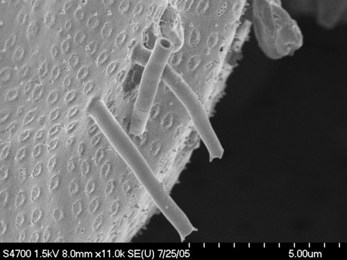
Cells quadrangular, connected in zigzag or straight chains attached at the elevations. Length of apical axis 10-97 µm. Valves are elliptical-lanceolate, with two obtuse elevations, and at each pole ringed ocelli having a dense pore field. Elevations are inflated at the base and divergent. The center part of valve is convex, more or less flattened at the top from which more or less long spine-like tubular labiate processes, two or many emerge. Frustule walls are heavy silicified, with valve areolae 8–11 in 10μ on the valve in radial rows. Girdle zone sharply differentiated from the valve zone by an expanded hyaline valve margin with an upturned rim. Areolae on the girdle band in pervalvar rows, 7–15 in 10 μm. Chloroplasts numerous, plate like and elliptical or reniform. Exhibits variations in morphology. (Cupp 1943; Hendey 1964; Hasle and Syvertsen 1996; Hoppenrath 2009; Plinksi and Witkowski 2020; An et al. 2001; Guiry and Guiry 2024)
Salish Sea specimens: Odentella aurita_Zm MHMPP_Mar 7-2021_BOX 1B T32_4 MQ-Mar 15-2022_m017_2.tif; Odontella aurita on Zm Mar 7-2021_T31 1Pb+c SA1b Stub2_s4800 RR_Oct 13-2021_m004_3.tif; Odontella on Zm Mar 7-2021_T31 1Pb+c SA1b Stub2_s4800 RR_Oct 13-2021_m005_3.tif; Odontella aurita Zm MHMPP Box 1b Stub 32-7 16 Mar 2022_b-RR_m013_4.tif; Odontella aurita- on Zm-H2O2 slide #1_4-Naphrax-E800-MU2003-Aug 4-2020-(Oct 23-2020-FS0023-0027_4.tif; Odontella aurita-T57 9D_B-HOT H2O2-TM4000 before 10Kv mode 3 H cushion(x1.5k)-July 23-2021 ECH_2_4.tif; Odontella aurita -SHW-IV-s4700- MW-July 14-05 m01-2b_4.tif; Odontella aurita -SHW-IV-s4700- MW-July 14-2005_ m10-4b.tif; Odontella aurita -SHW-IV-s4700- MW-July 14-2005_ m09-2b_3.tif; Odontella aurita -SHW-IV-s4700- MW-July 14-2005_ m02-4b.tif.
Morphometric data:
Cells quadrangular, connected in zigzag or straight chains. Length of apical axis 15.3-43.4 µm. Valves are elliptical-lanceolate, with obtuse elevations, ending in a ringed ocelli with a dense pore field inflated at the base. Ocelli elliptical length of 4-4.3 µm and a poroid density of 70-92 in 10 µm. The center part of valve is convex, more or less flattened at the top from which emerge more or less long external labiate spines, often two, sometimes several. Frustule walls are heavy silicified, with valve areolae 9-16 in 10μ on the valve in radial rows. Very short spinules on valve face. Girdle zone sharply differentiated from the valve zone by a clear depression. Areolae on the girdle band in pervalvar rows, 7-16 rows in 10 μm. Chloroplasts numerous, plate like and reniform. Exhibits considerable variations in morphology. Good support from molecular data for samples taken from SHW in 2022
Methods:
Odontella cells were collected in plankton net samples at Spanish Hills Wharf, Trincomali Channel, north Galiano Island.
Diatoms were collected by brushing or making razor blade scrapings of proximal, medial and distal sections. Additionally, 8-10 mm preserved leaf sections were cleaned with concentrated hydrogen peroxide or nitric acid at 100 C for 3-5 hours to remove organics, then rinsed multiple times in ddH20 to a neutral pH. Mounted on SEM stubs or in Naphrax on slides for light microscopy. Imaging with a Nikon TE300 and Tuscen DigiRetna16 MP camera or Nikon E800.
Live specimens were imaged with either a Nikon TE300 or Nikon E800 with either bright-field or DIC. In-situ, environmentally prepared samples were made using minimal contact of 8-10 mm leaf section, soaked in ddH2O to remove salts and dried through an EtOH series (50%-100%) and finished off with 100% Hexamethyldisilane HMDS (Hazrin-Chong and Manefield 2012). Mounted on carbon stickies onto aluminum SEM stubs and imaged with either the Hitachi s4800 or TM4000 at AMF, at University of Victoria, B.C. My thanks to Siobhan Schenk and Laura Parfrey in the Parfrey Lab at UBC for molecular data from the eelgrass and SHW samples, and collaboration with IMERSS. Also, thanks go to Elaine Humphrey of the AMF, UVIC, imaging by Ron Read, Melanie Quenneville and Arjan van Asselt. Additional imaging, taxonomy and identifications by Mark Webber (imerss.org).
References:
An, S.M.; Cho, K.; Kim, E.S.; Ki, H.; Choi, G.; Kang, N.S. (2023). Description and Characterization of the Odontella aurita OAOSH22, a Marine Diatom Rich in Eicosapentaenoic Acid and Fucoxanthin, Isolated from Osan Harbor, Korea. Mar. Drugs, 21,563. https://doi.org/10.3390/md21110563
Ashworth, M. P., Nakov, T., and Theriot, E. C. (2013). Revisiting Ross and Sims (1971): toward a molecular phylogeny of the Biddulphiaceae and Eupodiscaceae (Bacillariophyceae). Journal of Phycology, 49(6), 1207–1222. https://doi.org/10.1111/JPY.12131
Bérard-Therriault, L., Poulin, M. & Bossé, L. (1999). Guide d'identification du phytoplancton marin de l'estuaire et du Golfe du Saint-Laurent incluant également certains protozoaires. Publication Spéciale Canadienne des Sciences Halieutiques et Aquatiques 128: 1-387.
BOLD Systems: Taxonomy Browser - Odontella aurita {species} (February 13, 2021. https://v3.boldsystems.org/index.php/Taxbrowser_Taxonpage?taxid=87619, http://www.boldsystems.org/index.php/Public_RecordView?processid=DITS121-08
Cupp, E. E. (1943). Marine Plankton Diatoms of the West Coast of North America. Bull. Scrips. Inst. Oceanography. 5: 1-238.
Guiry, M.D. in Guiry, M.D. & Guiry, G.M. 2024. AlgaeBase. World-wide electronic publication, National University of Ireland, Galway. http://www.algaebase.org; searched on March 15, 2024.
Hasle, G.R. & Syvertsen, E.E. (1996). Marine diatoms. In: Identifying Marine Phytoplankton. (Tomas, C.R. Eds), pp. 5-385. San Diego: Academic Press.
Hazrin-Chong NH, Manefield M. (2012 ). An alternative SEM drying method using hexamethyldisilazane (HMDS) for microbial cell attachment studies on sub-bituminous coal. J Microbiol Methods. 90(2):96-9. doi: 10.1016/j.mimet.2012.04.014.
Hendey, N.I. (1964). An introductory account of the smaller algae of British coastal waters. Part V: Bacillariophyceae (diatoms). pp. [i]-xxii, 1-317. London: Ministry of Agriculture, Fisheries and Food, Fishery Investigations. Her Majesty’s Stationery Office.
Hoban, M.A. (1983). Biddulphioid diatoms II: The morphology and systematics of the Pseudocellate species, Biddulphia biddulphiana (Smith) Boyer, B. alternans (Bailey) Van Heurck, and Trigonium arcticum (Brightwell) Cleve. Botanica Marina, 26(6): 271-284
Hoppenrath, M., Elbrachter, M., Drebes, G. (2009) Marine Phytoplankton, Selected microphytoplankton species from the North Sea around Helgoland and Sylt. E. Schweizerbart’sche Verlagsbunchhandlung, Stuttgart, Germany.
Kützing, F.T. (1844). Die Kieselschaligen Bacillarien oder Diatomeen. pp. [i-vii], [1]-152, pls 1-30. Nordhausen: zu finden bei W. Köhne.
Lavigne, A.S., Sunesen, I. & Sar, E.A. (2015). Morphological, taxonomic and nomenclatural analysis of species of Odontella, Trieres and Zygoceros (Triceratiaceae, Bacillariophyta) from Anegada Bay (Province of Buenos Aires, Argentina). Diatom Research 30(4): 307-331.
Pienitz, R., Fedje, D. and Poulin, M. (2003) Marine and Non-Marine Diatoms from the Haida Gwaii Archipelago and Surrounding Coasts, Northeastern Pacific, Canada In Bibliotheca Diatomologica (H. Lange-Bertalot and P. Kociolek, eds.), Band 48, J. Cramer, Stuttgart, 146 pp.
McIntire, C. D. and Overton, W. S. (1971). Distributional Patterns in Assemblages of Attached Diatoms from Yaquina Estuary, Oregon. Ecology, Vol. 52, No. 5. pp. 758-777.
Rao, V.N.R. and Levin, J. 1976. Benthic marine diatom flora of False Bay, San Juan Island, Washington. Syesis, 9:173–213.
Round, F.E., Crawford, R.M. and Mann, D.G. (1990), The Diatoms, Biology & Morphology of the Genera, pp. 220-221. Cambridge University Press, Cambridge, UK.
Sancetta, C. and Calvert, S. E. (1988). The annual cycle of sedimentation in Saanich inlet, British Columbia: implications for the interpretation of diatom fossil assemblages. Deep Sea Research Part A. Oceanographic Research Papers, 35(1), 71–90. doi:10.1016/0198-0149(88)90058-1
Shim, J. H. (1976). Distribution and Taxonomy of Planktonic Marine Diatoms in the Strait of Georgia, B.C. Phd. Thesis, UBC.
Sims, P.A. (ed.) (1996). An atlas of British diatoms arranged by B. Hartley based on illustrations by H.G. Barber and J.R. Carter. pp. 406-409, Bristol: Biopress Ltd.
Sims, P. A., Williams, D. and Ashworth, M. P. (2018). Examination of type specimens for the genera Odontella and Zygoceros (Bacillariophyceae) with evidence for the new family Odontellaceae and a description of three new genera. Phytotaxa 382(1):1. DOI: 10.11646/phytotaxa.382.1.1
Spaulding, S.A., Bishop, I.W., Edlund, M.B., Lee, S., Furey, P., Jovanovska, E. and Potapova, M. Diatoms of North America. Retrieved November 6, 2021, from https://diatoms.org
Tynni, R. (1986). Observations of diatoms on the coast of the State of Washington. Geological Survey of Finland. Report of Investigation 75
Waters, R. E., Brown, L.N., and MG Robinson, M.G. (1992). Phytoplankton of Esquimalt Lagoon, British Columbia: comparison with west Vancouver Island coastal and offshore waters. Canadian Technical Report of Hydrography Ocean Sciences 137.